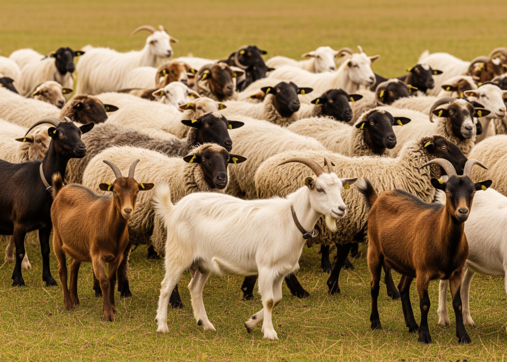
What Is Johne’s Disease in Cattle?
Johne’s disease in cattle, also known as paratuberculosis, is a long-term infectious condition that affects the digestive system of cattle and other ruminants. It is considered one of the important cattle bacterial diseases that can gradually impact herd health and productivity. The disease is caused by the bacterium Mycobacterium avium subspecies paratuberculosis, which mainly targets the lining of the small intestine.
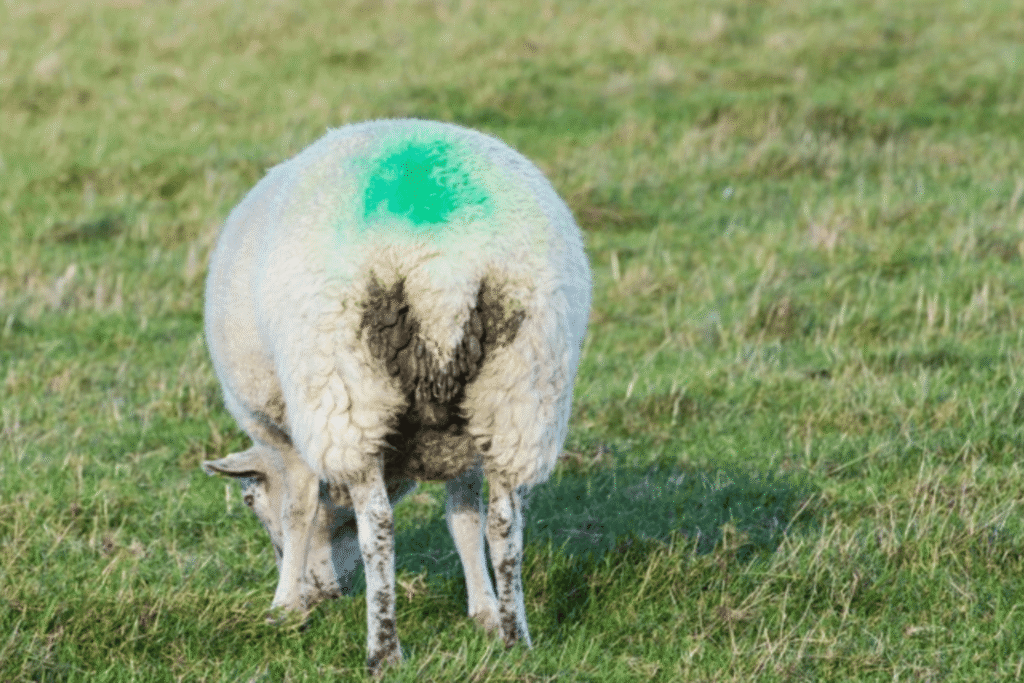
Over time, the infection damages the intestinal wall and reduces the animal’s ability to absorb nutrients properly. Because of this, affected cattle may start to lose weight even though they continue eating normally. Persistent diarrhea and a gradual decline in body condition are also common signs of this paratuberculosis disease.
Another reason Johne’s disease is difficult to manage is its slow development. In many cases, infected animals can carry the bacteria for years before clear symptoms appear. During this time, the infection may spread within the herd without being noticed, which makes early awareness and proper herd management essential for controlling paratuberculosis.
What is the Cause of Johne’s Disease in Cattle?
Johne’s disease in cattle is caused by the bacterium Mycobacterium avium subspecies paratuberculosis (MAP). This microorganism is responsible for paratuberculosis, a long-term infection that mainly affects the lower part of the small intestine in cattle and other ruminants.
One reason this disease develops slowly is that MAP can survive inside immune cells called macrophages. By living inside these cells, the bacteria can remain in the body for a long time while gradually damaging the intestinal tissues.
Another important feature of the bacteria that cause paratuberculosis disease is their ability to survive in the environment. Infected animals release large numbers of MAP bacteria in their feces. Smaller amounts of the bacteria may also be found in colostrum and milk. Once the bacteria enter the environment, they can remain alive for long periods. Studies show that MAP can survive for more than a year on pasture and may persist even longer in water than in soil.
After entering the body, the bacteria mainly settle in the Peyer’s patches located in the lower small intestine. Here, they infect macrophages in the intestinal tissue and nearby lymph nodes. The bacteria multiply slowly and cause long-term inflammation of the intestinal wall. Over time, this inflammation interferes with the intestine’s ability to absorb nutrients properly. Because the bacteria grow so slowly, Johne’s disease in cattle often takes several years before clear symptoms begin to appear.
Clinical Signs and Symptoms of Johne’s Disease in Cattle
The symptoms of Johne’s disease in cattle are Weight loss, diarrhea, and a severe case of bottle jaw, the weight loss even when cattle eat properly. Over time, affected animals develop cattle diarrhea, which may be persistent or intermittent. Because of this, farmers often notice cows with diarrhea within the herd.
Another important clinical sign is reduced productivity. In dairy cattle, milk production may drop or fail to reach expected levels. The hair coat may also appear dull or faded, which reflects poor nutrient absorption and declining body condition.
In advanced cases, animals may develop swelling under the jaw known as bottle jaw in cattle or bottle jaw cow. This occurs due to protein loss caused by intestinal damage. As the disease progresses, animals may become severely thin and weak, and carcasses can appear emaciated due to long-term nutrient loss.
These signs can resemble other cattle disease symptoms, such as the symptoms of worms in cattle that also cause diarrhea and weight loss. However, the combination of chronic diarrhea, progressive weight loss, reduced milk yield, and bottle jaw in cattle are key symptom of Johne’s disease in cattle.
Johne’s disease symptoms usually appear in the later stages of infection and mainly affect the digestive system. One of the most common symptoms of Johne’s disease in cattle is progressive weight loss, even when the animal continues to eat normally. In advanced cases, the animal may become extremely thin, and carcasses may appear emaciated with visible ribs and bones.
Another key sign is chronic cattle diarrhea. Many infected animals are first noticed as a cow with diarrhea that continues for a long time. The diarrhea may be constant or intermittent, and farmers may observe cows with diarrhea that gradually lose body condition.
As the disease progresses, the coat may become rough or faded, and milk production may drop or fail to reach expected levels, especially in dairy cattle. In severe cases, affected animals may develop bottle jaw in cattle, a swelling under the jaw caused by protein loss from the damaged intestine. This condition is often referred to as a bottle jaw cow.
Some of these signs can also appear in other conditions. For example, heavy parasitic infections may produce symptoms of worms in cattle, including diarrhea, weight loss, and swelling under the jaw. Recognizing these cattle disease symptoms early helps farmers and veterinarians manage the disease and prevent its spread within the herd.
How Johne’s Disease Spreads in Cattle
Johne’s disease, which is caused by bacteria Mycobacterium avium subspecies paratuberculosis (MAP), can be transferred through Fecal–Oral Route. This bacterium causes Paratuberculosis, a long-term infection that mainly affects the intestines and moves slowly through a herd. The bacteria are mostly passed in the manure of infected animals. From there, they can easily contaminate feed, drinking water, milk, and even the general farm environment.
(Fecal–Oral Route)
In simple terms, the main way this disease spreads is when cattle accidentally eat or drink something contaminated with infected manure.
This usually happens when:
- Feed or water gets dirty with manure
- Calves are kept in areas that aren’t cleaned properly.
- Animals graze on land where infected cattle have been
One tricky thing about MAP is that it can survive in the environment for quite a long time, so once an area is contaminated, it can keep infecting animals if not managed properly.
Milk Feeding
Young calves are the most at risk. In many cases, calves become infected very early in life, but farmers often don’t notice any signs until the animals are older.
This can happen when:
- A calf ingests manure soon after birth
- It drinks infected milk or colostrum
- It stays in contaminated bedding or calving areas
Sometimes, the infection can even pass from the mother to the calf during pregnancy, though this is less common.
The Role of the Environment
The environment plays a big part in spreading the disease. MAP bacteria can live for months in soil, water, and manure.
Common sources of infection around the farm include:
- Feed and water troughs contaminated with manure
- Wet, muddy, or poorly maintained housing
- Pastures that were previously used by infected cattle
If hygiene isn’t maintained, the bacteria can quietly build up and keep spreading within the herd.
Spread Through Milk and Colostrum
Milk can also be a source of infection, especially for young calves.
This risk increases when:
- Calves are given raw milk from infected cows
- Colostrum from different cows is mixed.
- Milk gets contaminated with manure during milking.
Good milking hygiene and careful feeding practices can go a long way in reducing this risk.
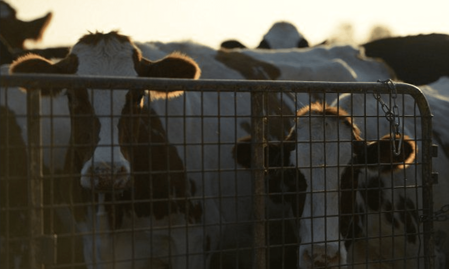
Bringing Infection Into the Herd
The disease often spreads when farmers introduce new animals that are already infected, even if they appear healthy.
This can happen when:
- Cattle are bought from unknown or infected farms
- New animals are added to the herd without testing or quarantine
These animals may not show signs right away, but they can still spread Paratuberculosis over time.
Diagnosis of Johne’s Disease in Cattle
Diagnosing Johne’s disease in cattle is mainly based on laboratory testing combined with clinical observation. Because Johne’s infection progresses slowly, many animals remain infected for a long time before showing clear symptoms. Therefore, veterinarians confirm the disease by detecting the bacteria, identifying the immune response, or examining affected tissues.
Clinical Evaluation
The diagnosing process starts by observing animals that show signs such as chronic diarrhea, weight loss, and reduced milk production. These symptoms typically appear in the later stages of Johne’s disease.
The disease is commonly described in four stages:
- Stage 1: Infected animals show no symptoms, and bacteria are not detected in feces.
- Stage 2: Animals remain asymptomatic but may begin shedding bacteria in feces.
- Stage 3: Clinical signs such as diarrhea and weight loss appear.
- Stage 4: Advanced disease with severe emaciation, weakness, and persistent diarrhea.
Detection of the Bacteria
Veterinarians confirm Johne’s disease by detecting MAP bacteria in fecal or tissue samples.
Common methods include:
- Acid-fast staining (Ziehl–Neelsen stain) to identify mycobacteria in samples
- Bacteriologic culture to grow MAP bacteria from feces or tissues.
- Pooled fecal culture for screening multiple animals within a herd
Culture methods are reliable but may take several weeks due to slow bacterial growth.
PCR Testing
Veterinarians widely use PCR (Polymerase Chain Reaction) as a diagnostic tool for Johne’s disease. It detects the DNA of MAP bacteria in feces, milk, or tissue samples and provides faster results than traditional culture methods.
Immunologic and Serologic Tests
Diagnosis may also involve tests that detect the immune response of cattle to Johnes infection.
These include:
- Skin testing for delayed-type hypersensitivity (DTH)
- Gamma interferon (IFN-γ) tests
- Serologic tests such as ELISA, AGID, and Complement Fixation
Among these, ELISA is commonly used for herd screening because it is quick and cost-effective.
Tissue Examination
In advanced cases of Johne’s disease, veterinarians may examine intestinal tissues, especially from the ileum and regional lymph nodes. Veterinarians can test tissue samples using PCR, culture, or acid-fast staining, which may reveal large numbers of mycobacteria in intestinal lesions.
Confirming the Disease
A confirmed diagnosis of Johne’s disease usually involves:
- Detecting MAP bacteria in feces or tissues using PCR or culture
- Identifying antibodies against MAP through blood tests such as ELISA
- Observing characteristic intestinal lesions during pathological examination
Using multiple diagnostic methods helps accurately identify cattle infected with Johnes and supports effective herd management.
Treatment for Johne’s Disease in Cattle
There is no treatment available for Johne’s Disease in cattle. Management, like supportive care, may work to cure the cattle. The disease is caused by Mycobacterium avium subspecies paratuberculosis (MAP), a bacterium that damages the lining of the small intestine. As the infection progresses, affected cattle develop chronic diarrhea, gradual weight loss, and poor nutrient absorption.
Because the bacteria live deep inside intestinal tissues, eliminating the infection is very difficult. For this reason, Johne’s disease treatment mainly aims to manage symptoms, improve the animal’s comfort, and maintain body condition.
Nutritional Support
Proper nutrition is important in managing Johne’s disease. Infected cattle often lose weight because their intestines cannot absorb nutrients efficiently. Providing high-energy and easily digestible feed can help slow weight loss and support the animal’s condition.
Several supportive supplements are commonly used in cow disease treatment programs:
Propylene glycol – helps improve energy balance in weak or underweight animals.
Dose: 250–400 ml orally once daily for 3–5 days.
Vitamin injections (AD3E or Vitamin B-complex) – support metabolism and may improve appetite.
Dose: 5–10 ml intramuscularly, depending on the product formulation.
Probiotics or rumen stimulants – yeast-based supplements that help improve rumen activity and digestion.
Dose: 20–40 g orally per day mixed with feed.
These products help support the animal’s condition but do not cure Johne’s disease.
Fluid and Electrolyte Therapy
Dehydration is a common risk in cattle with Johne’s disease because chronic diarrhea causes loss of fluids and minerals.
Fluid and electrolyte therapy is therefore often included in treatment.
Common options include:
Oral electrolyte solutions – Restores fluids and minerals lost through diarrhea.
Dose: 2–5 liters per day, depending on dehydration level.
Intravenous fluids such as Ringer’s lactate or normal saline are used in severe cases under veterinary supervision.
Dose: 5–10 liters IV when dehydration is significant.
Maintaining hydration helps stabilize affected animals and improves their overall condition.
Anti-Diarrheal Support
Managing chronic diarrhea is another important part of Johne’s disease treatment. Persistent diarrhea can irritate the intestinal lining and weaken the animal.
Certain intestinal protectants and supportive medications may help reduce irritation and fluid loss.
Commonly used products include:
Kaolin-pectin mixtures – help coat the intestinal lining and reduce diarrhea.
Dose: 100–200 ml orally twice daily.
Activated charcoal or toxin binders – sometimes used to support intestinal health and absorb harmful substances in the digestive tract.
These treatments mainly help control symptoms and support digestive health.
Antibiotic Therapy for Johne’s Disease in Cattle.
Although Johne’s disease is bacterial, antibiotics are generally not effective for routine treatment in cattle. Drugs such as rifampin, clarithromycin, and clofazimine have been studied against MAP.
However, they require long-term use, can be expensive, and are usually not practical for most cattle farms. Veterinarians rarely recommend antibiotics as a standard treatment for this disease.
Advanced Clinical Cases
In later stages of Johne’s disease, cattle may show severe symptoms such as:
- Extreme weight loss
- Persistent watery diarrhea
- Weakness and dehydration
At this stage, intestinal damage is often extensive, and treatment usually provides only temporary relief. Management on farms, therefore, often focuses on preventing disease spread and identifying infected animals early.
Prevention of Johne’s Disease in Cattle
Preventing Johne’s disease in cattle mainly relies on strict hygiene and limiting exposure to Mycobacterium avium subspecies paratuberculosis (MAP). Separate calves immediately after birth, fed clean colostrum, and raised in a dry, manure-free area to avoid early exposure.
Keep their feed, water, barns, and calving spaces clean and well-managed. Vaccines such as Gudair and Silirum can also help control the spread. When bringing in new animals, it’s best to source them from reliable, disease-managed herds and keep them in quarantine before mixing them with the rest.
Protect Young Calves From Infection
Young calves are much more vulnerable to Johne’s disease than adult cattle. In many cases, animals become infected early in life but only show symptoms years later. For this reason, protecting calves during the first months of life is one of the most important steps in preventing paratuberculosis disease.
Some simple but effective practices include:
- Separating the calf from the cow soon after birth.
- Keeping calves in clean and dry housing areas that are free from manure contamination.
- Feeding clean colostrum from healthy cows.
- Avoiding pooled milk or colostrum from animals with unknown health status.
These steps help reduce the chances of calves becoming infected with Johnes in cattle at an early age.
Maintain Farm Hygiene
The bacteria that cause Johne’s disease mainly spread through manure. When manure contaminates feed, water, or bedding, other animals can easily ingest the bacteria. Maintaining good hygiene on the farm, therefore, plays a major role in cattle disease prevention.
Important hygiene practices include:
- Keeping feed bunks and water troughs free from manure.
- Regularly cleaning barn floors, feeding areas, and calving pens.
- Avoid spreading manure from infected cattle on pastures where calves graze.
- Use clean bedding and replace it frequently.
Consistent sanitation helps lower the number of bacteria in the environment and reduces the spread of bacterial diseases in cattle, including Johne’s disease.
Vaccination for Johne’s Disease
Vaccination can also help control Johne’s disease in herds where the infection is already present. While vaccines cannot completely prevent infection, they may reduce the severity of disease and decrease the number of bacteria shed by infected animals.
Some commonly used vaccines include:
- Gudair® Vaccine
- Dose: 1 ml given as a subcutaneous injection, usually once to calves between 1–4 months of age.
- Silirum® Vaccine
- Dose: 1 ml subcutaneous injection, typically administered once in young calves.
A veterinarian should always supervise vaccination, as improper administration can cause local reactions.
Isolation of New Animals
One of the most common ways Johne’s disease enters a herd is through the purchase of infected animals. Even cattle that look healthy may carry the bacteria and spread them to others.
To reduce this risk:
- Buy cattle only from trusted herds with good disease management.
- Avoid animals showing chronic diarrhea or unexplained weight loss.
- Keep newly purchased cattle separate from the main herd for a period of observation.
These precautions help prevent the introduction of paratuberculosis disease and support overall cattle disease prevention on the farm.
Conclusion
Johne’s disease in cattle is a gradually-developing bacterial infection caused by Mycobacterium avium subspecies paratuberculosis (MAP) that mainly affects the digestive system and can gradually reduce herd productivity. Infected animals may carry the bacteria for several years before clear signs appear, such as weight loss, long-lasting diarrhea, reduced milk production, and bottle jaw.
Veterinarians usually confirm the disease through laboratory tests like PCR, bacterial culture, or blood tests. Because there is no reliable cure for Johne’s disease, treatment mainly focuses on supportive care to help affected animals maintain their condition.
Prevention is the most practical approach. Protecting young calves, Feeding and housing areas should be clean, managing milk and colostrum carefully, vaccinating where appropriate, and introducing new animals only from well-managed herds can help reduce the spread of Johne’s disease and protect the overall health of the herd.
FAQ,s related to Johne’s disease in cattle
What is Johne’s disease in cows?
Johne’s disease in cows is a chronic bacterial infection that affects the intestines, caused by Mycobacterium avium subspecies paratuberculosis (MAP). It slowly reduces nutrient absorption, leading to weight loss, diarrhea, and lower milk production over time.
How common is Johne’s disease in cattle?
Johne’s disease is fairly common in cattle herds worldwide, especially in dairy farms. Many infected animals carry the bacteria for years without showing symptoms, which makes it easy for the disease to spread unnoticed.
How to test for Johne’s disease in cattle?
Veterinarians usually test cattle for Johne’s disease using lab methods like PCR, fecal culture, or blood tests (ELISA) to detect the bacteria or the animal’s immune response. Vets often combine these tests with observation of clinical signs for accurate diagnosis.
What are the symptoms of Johne’s disease in cattle?
The main symptoms of Johne’s disease in cattle are chronic diarrhea, gradual weight loss despite normal appetite, reduced milk production, and, in severe cases, swelling under the jaw (bottle jaw). Early signs can be subtle, making the disease hard to detect.